
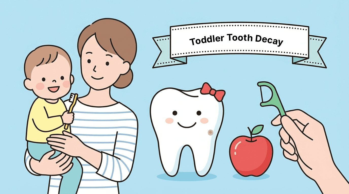
Toddler-Tooth-Decay-Causes,-Early-Signs-How-to-Prevent-Cavities-in-Young-Kids
Toddler Tooth Decay: Causes, Early Signs & How to Prevent Cavities in Young Kids
Toddler tooth decay is the most common chronic disease in children worldwide — more prevalent than asthma, more widespread than childhood obesity, and yet among the most preventable conditions in all of medicine.
The dental chair is cartoonishly oversized for a two-year-old. There are stickers on the ceiling lights — a smiling giraffe, a rocket ship — and the hygienist is wearing a pair of gloves patterned with tiny dinosaurs. Everything in the room has been designed to telegraph safety, warmth, friendliness. And yet the parent sitting six feet away, watching the dentist gently retract their toddler’s upper lip, feels a slow chill settle in their chest. Because there, along the gumline of those impossibly small front teeth, are white chalky patches — or, in more advanced cases, brown crumbling craters where firm, healthy enamel once stood.
How did I miss this?
That question — whispered, thought, sometimes wept — echoes through pediatric dental offices thousands of times every single day. And what makes it so particularly devastating is the shame that rides alongside it: the unspoken belief that a cavity in a toddler’s mouth is a verdict on one’s parenting, a scarlet letter etched in enamel.
It is not. What it is, however, is extraordinarily common — and understanding why requires rethinking almost everything most people believe about tooth decay.
Early childhood caries (ECC), the clinical term for tooth decay in children under age six, is not a niche medical curiosity. According to the World Health Organization, it is the single most prevalent chronic disease in childhood globally, affecting an estimated 530 million children. To put that in perspective, the entire population of the European Union is roughly 450 million. In the United States alone, the Centers for Disease Control and Prevention reports that more than 20 percent of children aged two to five have at least one untreated cavity. That rate surpasses the prevalence of childhood asthma, a condition that commands enormous public health infrastructure, school nurse training programs, and billions in research funding.
Toddler tooth decay, by contrast, hides in plain sight. It is dismissed by a myth so pervasive it has become almost axiomatic: They’re just baby teeth — they’ll fall out anyway. That myth, as this article will demonstrate, is not merely inaccurate. It is one of the most costly misconceptions in all of pediatric medicine, and dismantling it begins with understanding what is actually happening inside a toddler’s mouth.
What Is Actually Happening Inside a Toddler’s Mouth
To understand tooth decay, it helps to first understand what a tooth actually is — because most people, understandably, have never had reason to think about teeth as anything other than hard white objects that chew food.
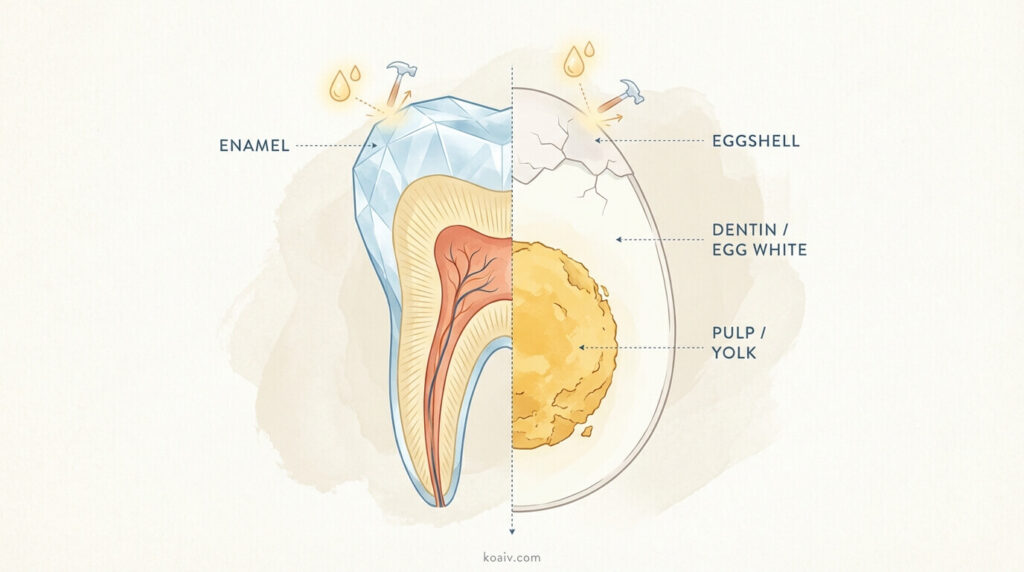
A tooth is, in fact, a living structure. Beneath the visible white exterior lies a layered architecture not unlike a hard-boiled egg. The outermost shell — the enamel — is the hardest substance in the human body, harder even than bone. It is a crystalline mineral matrix composed almost entirely of calcium and phosphate, and its job is to serve as armor: protecting the softer, more sensitive layers beneath from the mechanical forces of chewing and, critically, from the chemical assault of acid.
Beneath the enamel sits the dentin, a yellowish, porous layer that is considerably softer and more vulnerable. Think of it as the egg white — structurally important but defenseless without the shell above it. And at the very center of the tooth lies the pulp, the egg yolk equivalent: a chamber of living nerve tissue and blood vessels that, when breached by decay, triggers the throbbing, debilitating pain any adult with a deep cavity will recognize instantly.
Here is the detail that changes the entire calculus for toddlers: baby teeth have dramatically thinner enamel than adult teeth. Where a permanent molar might wear a suit of armor two to three millimeters thick, a primary incisor may have less than one millimeter of enamel protecting its nerve.
If the enamel of an adult tooth is a castle wall, the enamel of a baby tooth is a garden fence. The protective barrier is real, but it is far more easily breached — and once it is, decay reaches the sensitive inner layers with startling speed. A cavity that might take a year or more to become problematic in an adult tooth can reach the nerve of a baby tooth in a matter of weeks.
This structural vulnerability is not a design flaw. Baby teeth were never engineered to last a lifetime. They are temporary structures built for a temporary purpose — holding space, facilitating speech, enabling nutrition during the years when the jaw is too small for permanent teeth. But the fact that they are temporary does not make them disposable, a distinction that carries enormous consequences and one that deserves its own dedicated discussion later in this article.
The Acid Attack: Decay as a Chemical Process
Understanding what decay actually is — at a molecular level — strips away much of its mystery and, more importantly, reveals exactly where the leverage points for prevention lie.
Tooth decay is not, as many intuitively assume, a process of sugar somehow “eating” through the tooth. It is a chemical reaction mediated by bacteria, and it works like this: hundreds of species of bacteria live in the human mouth at any given time, forming a sticky, transparent film on the tooth surface called plaque. (That slightly fuzzy feeling on the teeth after a day without brushing? That is plaque.) Among these hundreds of species, a subset — most notably a bacterium called Streptococcus mutans — has a particular metabolic talent. When these bacteria encounter fermentable carbohydrates (sugars, starches, anything that breaks down into simple sugars), they consume them and excrete lactic acid as a metabolic byproduct.
That acid is the weapon. It lowers the pH on the tooth surface — in other words, it makes the local environment more acidic — and at a certain threshold (roughly pH 5.5), the crystalline mineral structure of the enamel begins to dissolve. Calcium and phosphate ions leach out of the enamel surface in a process scientists call demineralization. Imagine a limestone cliff face being slowly eroded by acidic rain — the dynamic is chemically identical.
But here is the hopeful part of the equation: this process is not a one-way street. Saliva— that underappreciated, constantly flowing fluid — acts as the mouth’s built-in repair crew. It is rich in calcium, phosphate, and bicarbonate buffers. After an acid attack subsides (typically when the carbohydrate source is consumed or cleared away), saliva gradually neutralizes the acid, raises the pH back to safe levels, and begins depositing minerals back into the weakened enamel surface. Scientists call this counter-process remineralization. Think of it as the tide coming back in after an unusually low ebb: the beach is restored, the damage undone, as long as the recovery period is long enough.
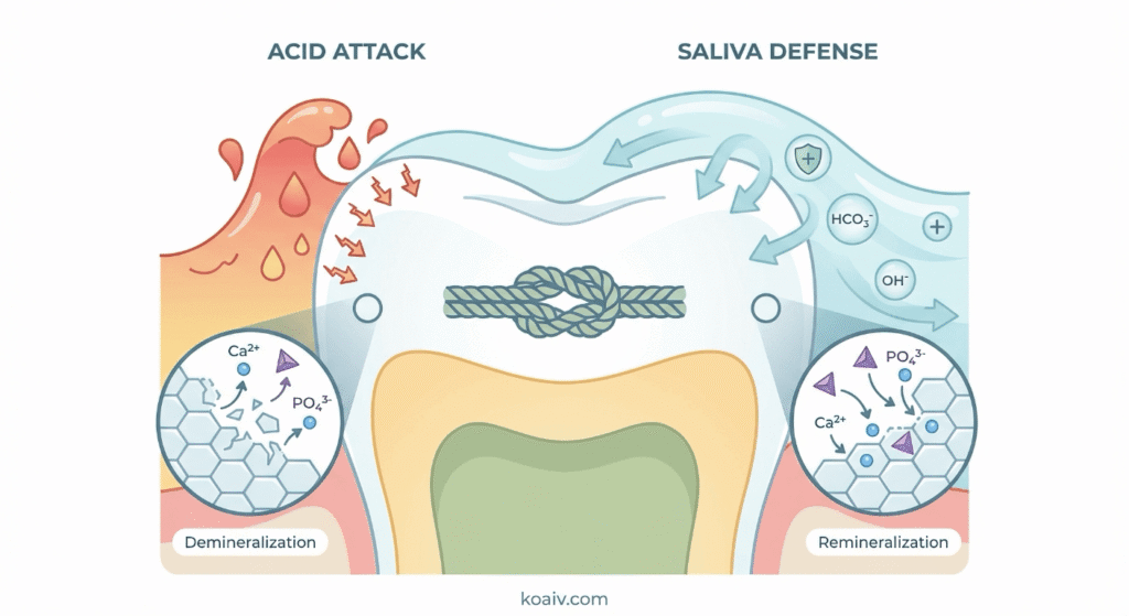
The entire battle for a toddler’s teeth, then, can be understood as a tug-of-war between demineralization and remineralization — between acid attacks and saliva recovery. When the recovery periods win, the teeth hold. When the acid attacks come too frequently, too relentlessly, with too little recovery time between them, the enamel crumbles. Everything that follows in this article — the dietary advice, the brushing protocols, the fluoride recommendations — is ultimately about tilting that balance decisively in favor of remineralization.
Why Toddlers Get Cavities
If tooth decay is a chemical war between acid and repair, then it stands to reason that the causes of cavities are anything that either amplifies the acid attacks or undermines the repair process. In toddlers, these causes form a constellation of factors — some obvious, some deeply counterintuitive, and some that challenge fundamental assumptions about what it means for a disease to be “preventable.”
Frequency Matters More Than Quantity
Of all the insights in pediatric dentistry, this one may be the most important for parents to internalize — and it is also the most counterintuitive.
Most people assume that the total amount of sugar a child consumes in a day determines cavity risk. Eat less sugar, get fewer cavities. This is, at best, a half-truth. What research has conclusively demonstrated is that the frequency of sugar exposure matters far more than the total volume. The reason traces directly back to the acid-attack cycle described above.
Every time a carbohydrate enters the mouth — every sip of juice, every nibble of a cracker, every handful of cereal puffs — the bacteria in plaque begin producing acid almost immediately. The pH on the tooth surface drops into the danger zone within three to five minutes and stays there for roughly 30 to 45 minutes before saliva can buffer it back to safety. This means that a single snacking event triggers a single acid attack lasting about half an hour. The teeth experience one assault, followed by recovery.
Now consider a toddler who carries a sippy cup of diluted apple juice throughout the morning, taking a small sip every ten or fifteen minutes. Each sip — no matter how small — resets the clock. The pH drops, the 30-minute timer restarts, the enamel never gets its recovery window. Over the course of a three-hour morning, the teeth may be under continuous acid siege without a single minute of remineralization. The total volume of juice consumed might be quite modest — perhaps four ounces — but the pattern of exposure is catastrophic.
By contrast, a child who eats an entire cookie at a designated snack time, drinks a glass of water, and then doesn’t eat again for two hours experiences a single, time-limited acid attack followed by a full recovery period. The teeth rebuild. The balance tips toward health.
This is the fundamental principle behind every dietary recommendation in pediatric dentistry: structured eating patterns with clear breaks between meals. It is not about deprivation. It is about giving the mouth time to heal between exposures.
The Bedtime Bottle: A Perfect Storm
One behavioral pattern crystallizes this frequency principle into its most dangerous form: putting a child to sleep with a bottle of milk, formula, or juice.
During sleep, saliva production drops to its lowest point — a natural circadian rhythm that, under normal circumstances, causes no harm. But when a child falls asleep with a bottle, the liquid pools around the upper front teeth (which are positioned directly behind the upper lip, exactly where the bottle nipple rests). The teeth sit in a bath of lactose, sucrose, or fructose for hours, bacteria produce acid continuously, and saliva — the only defense — has essentially gone off duty.
The result is a pattern of decay so distinctive that dentists gave it its own name decades ago: “baby bottle tooth decay.” The upper front teeth erode in a characteristic arc along the gumline while the lower front teeth, partially shielded by the tongue and bathed more directly in the sublingual saliva glands’ output, often remain untouched.
The same dynamic applies to prolonged nighttime breastfeeding. While breast milk alone is less cariogenic (cavity-causing) than formula or juice due to its lower sugar concentration and the presence of protective antibodies, the mechanics of prolonged overnight nursing — pooling liquid, reduced saliva, uninterrupted bacterial feeding — still create conditions that can contribute to ECC, particularly once a full complement of front teeth has erupted.
Hidden Sugars and the Stickiness Factor
Parents tend to feel confident about identifying sugary foods: candy, cookies, soda, ice cream. These are the obvious suspects, and most caregivers already manage them with some degree of intentionality. What consistently catches parents off guard are the foods that seem healthy — or at least neutral — but are, from a dental perspective, surprisingly problematic.
Dried fruit is perhaps the most common offender. Raisins, dried cranberries, and dried mango are often included in toddler snack mixes precisely because they feel like wholesome, natural choices. And nutritionally, they have genuine virtues. But from a dental standpoint, they represent a near-perfect combination of concentrated sugar and extreme stickiness. A raisin can adhere to the deep grooves of a molar for hours, providing a sustained, slow-release sugar source directly to the tooth surface — an all-day buffet for S. mutans.
Crackers, puffs, and other refined carbohydrates are equally deceptive. They may not taste sweet, and many parents do not mentally categorize them as “sugary,” but the starches in white flour are rapidly broken down by salivary enzymes (specifically, amylase) into simple sugars within minutes of entering the mouth. A handful of goldfish crackers is, from the bacteria’s perspective, functionally identical to a handful of sugar cubes that dissolve slowly.
Gummy vitamins deserve particular mention because they represent a kind of conceptual whiplash for parents: a product purchased explicitly for health purposes that simultaneously increases cavity risk. Their chewy, sticky texture ensures prolonged contact with tooth surfaces, and their sugar content — necessary to make them palatable to children — feeds the very bacteria that undermine dental health.
Fruit pouches, those ubiquitous squeezable tubes that have become a staple of modern toddler nutrition, often contain more sugar per serving than a scoop of ice cream. Because the pouch is typically squeezed directly into the mouth, the sugary purée bypasses the natural dispersal that occurs when food is chewed and instead concentrates directly on specific tooth surfaces.
The Invisible Transmission: Cavities as an Infectious Disease
Here is where the conversation about toddler tooth decay departs most dramatically from the conventional understanding — and enters territory that, for many parents, is genuinely paradigm-shifting.
Tooth decay is, at its biological core, an infectious disease. The bacteria that cause it are not native residents of a newborn’s mouth. Babies are born with essentially sterile oral cavities — they harbor no Streptococcus mutans at birth. These cavity-causing bacteria must be acquired from an external source, and decades of microbiological research have established that the source is almost always a primary caregiver, most commonly a parent.
Scientists call this process vertical transmission, and it happens through entirely mundane, loving acts: tasting food from the same spoon before offering it to the baby, cleaning a fallen pacifier in one’s own mouth, sharing a cup, blowing on a bite of food to cool it down. A landmark 2008 study published in the Journal of the American Dental Association tracked mother-infant pairs and found that children whose mothers had high salivary concentrations of S. mutans were significantly more likely to develop early cavities — even after controlling for other risk factors like diet and hygiene.
This finding carries a profoundly practical implication: a caregiver’s own oral health directly influences the cavity risk of their child. A parent who addresses their own untreated cavities, maintains consistent brushing and flossing, and reduces their own bacterial load during pregnancy and the first years of a child’s life is, in a very real and measurable sense, lowering the child’s caries risk through a mechanism that has nothing to do with the child’s own teeth.
This is emphatically not information designed to produce guilt. It is information designed to widen the aperture of prevention — to show that the fight against ECC begins not in the nursery but in the parent’s own dental chair.
The Overlooked Factor: Mouth Breathing and the Erosion of Natural Protection
One risk factor that remains conspicuously absent from most popular discussions of childhood cavities is chronic mouth breathing. Its omission is significant because it affects a substantial minority of toddlers and because its contribution to decay risk is both well-documented and highly actionable.
Saliva, as established earlier, is the mouth’s natural defense against acid. It buffers pH, delivers remineralizing minerals to weakened enamel, and contains antimicrobial proteins that help keep bacterial populations in check. When a child chronically breathes through the mouth — whether due to enlarged adenoids, persistent allergies, a deviated septum, or habitual posture — the oral environment dries out, and saliva’s protective function is dramatically compromised.
The clinical consequences are measurable and consistent. Children who are chronic mouth breathers show higher rates of cavities, more plaque accumulation, and increased gingival (gum) inflammation compared to nasal breathers, even when all other variables — diet, hygiene, fluoride exposure — are held constant.
Parents who notice that a child habitually sleeps with the mouth open, snores regularly, or seems to breathe predominantly through the mouth during waking hours should bring this observation to both their pediatrician and their dentist. In many cases, the underlying obstruction — enlarged adenoids being the most common culprit — can be treated, and addressing it yields benefits that extend far beyond the dental realm and into improved sleep quality, behavioral regulation, and craniofacial development.
Why Baby Teeth Matter Far More Than Anyone Thinks
This is the section of the article that asks readers to set aside one of the most deeply held assumptions in popular understanding of dentistry — the belief that primary teeth, because they are temporary, are fundamentally unimportant. This belief is so intuitive, so logically tidy, that dislodging it requires more than a simple assertion to the contrary. It requires understanding the specific, tangible, and sometimes irreversible consequences that flow from untreated decay in primary teeth.
The Architecture of the Jaw: Baby Teeth as Space Holders
Permanent teeth do not erupt randomly. Each one follows a genetically determined path toward a specific location in the jaw, guided in part by the position of the primary tooth that precedes it. Baby teeth serve as natural space maintainers — biological placeholders that preserve the three-dimensional architecture the permanent teeth will eventually need.
When a baby molar is lost prematurely — whether to decay, infection, or extraction — the neighboring teeth begin to drift into the vacant space. This happens gradually, often imperceptibly, over weeks and months. By the time the permanent tooth is ready to erupt, years later, the doorway it was supposed to walk through has narrowed or closed entirely. The permanent tooth becomes impacted (trapped beneath the gumline) or erupts in a misaligned position, crowding its neighbors and distorting the bite.
The downstream consequence is orthodontic treatment — braces, expanders, sometimes surgical intervention — that can span years and cost tens of thousands of dollars. And while orthodontic correction is highly effective, not every family has access to it. The premature loss of a two-dollar baby tooth can set in motion a cascade of structural complications that reverberates through adolescence and into adulthood.
Speech and Language: The Hidden Connection
The upper front teeth — the very teeth most commonly devastated by baby-bottle decay — are not merely cosmetic features. They are active participants in speech production. Sounds like /s/, /z/, /f/, /v/, /sh/, and /th/ are articulated by directing airflow against or between the front teeth and the tongue or lip. A child who loses these teeth at age two or three — well before the permanent replacements arrive around age six or seven — must learn to compensate, often developing atypical tongue placements and compensatory speech patterns that, if established during the critical window of language acquisition, can become habitual and require formal speech therapy to correct.
The connection between dental health and speech development is one that parents rarely make intuitively, in part because the two domains — dentistry and speech-language pathology — are typically siloed in both clinical practice and public awareness. But for the toddler who has lost multiple front teeth to decay, the consequences are not theoretical. They are audible in every sentence.
Nutrition, Pain, and the Failure-to-Thrive Connection
A toddler with multiple active cavities is a toddler in pain — and dental pain in a child who cannot yet fully articulate the location, intensity, or nature of the discomfort manifests in ways that are easy to misattribute. Irritability, disrupted sleep, refusal of certain foods, clinginess, difficulty concentrating at preschool: all of these behaviors have dental pain as a potential (and frequently overlooked) root cause.
Children with untreated decay tend to modify their diets unconsciously, gravitating toward soft, easy-to-chew foods and avoiding anything crunchy, fibrous, or requiring sustained chewing — which, unfortunately, describes many of the most nutrient-dense foods available to young children. A 2007 study in Pediatric Dentistry found that children with severe early childhood caries weighed significantly less than their cavity-free peers and showed measurable deficits in height-for-age scores, a clinical marker associated with chronic undernutrition. The mechanism is straightforward: a child who avoids chewing because chewing hurts will consume fewer calories and fewer nutrients, and over time, growth falters.
When Decay Becomes an Emergency
Perhaps the most sobering consequence of untreated ECC is its potential to escalate from a dental problem into a medical emergency. A cavity that progresses through the enamel, through the dentin, and into the pulp creates an open pathway for bacteria to enter the bloodstream and the deeper tissues of the face and neck.
A periapical abscess — an accumulation of pus at the root tip of an infected tooth — can cause dramatic facial swelling, fever, difficulty swallowing, and, in rare but well-documented cases, spread to the orbital (eye) region or the deep fascial spaces of the neck, creating a life-threatening emergency. Pediatric emergency departments across the country treat children every week for dental infections that began as simple, treatable cavities and escalated through months or years of neglect. These cases are, without exception, preventable.
Toddler Tooth Decay Warning Signs
The pediatric dental community has worked to equip parents with a simple, zero-cost screening tool that can be performed at home and requires no equipment beyond a well-lit room and a gentle touch: the “Lift the Lip” check.
The technique is exactly what it sounds like. Once a month — during a bath, during a diaper change, during any quiet moment when the child is relaxed and the lighting is adequate — gently lift the child’s upper lip to fully expose the front surfaces and gumlines of the upper teeth. Look carefully. What appears there tells a remarkably clear story about the trajectory of the child’s oral health.
White chalky spots appearing along the gumline are the earliest visible sign of demineralization — areas where acid has begun to leach minerals from the enamel but has not yet broken through the surface. These spots are dull and opaque, lacking the healthy translucent sheen of intact enamel. If they look like someone dabbed white-out along the base of the teeth, that is cause for a prompt (but not panicked) dental visit.
This stage is critically important because it represents the only point in the decay process that is fully reversible without drilling. With professional fluoride application, improved oral hygiene, and dietary adjustment, remineralization can restore these areas to full structural integrity. This is the window every parent hopes to catch.
When those white spots progress — when the surface enamel weakens enough to allow staining and the underlying dentin becomes visible — yellow or light brown discoloration appears. The decay has now crossed the threshold from reversible to irreversible; the enamel has been breached, and professional treatment is needed to halt further progression. At this stage, treatment is still relatively conservative — a small filling, a fluoride varnish application, or in some cases Silver Diamine Fluoride (discussed below) — but the window for non-invasive remineralization has closed.
Further progression produces visible pitting, holes, and structural collapse — the tooth surface craters inward, and the decay becomes obvious to the naked eye. Treatment at this stage typically requires a more substantial restoration: a filling, a crown, or, if the nerve is involved, a procedure analogous to a root canal.
In the most advanced cases, the tooth may be reduced to a dark brown or black stump at the gumline, sometimes accompanied by a small, raised bump on the gum tissue that resembles a pimple. That bump — known clinically as a fistula or sinus tract— is the body’s attempt to drain an abscess forming at the root tip. It is not painful in many cases (the pressure is being relieved through the drainage path), which is precisely why it can go unnoticed for weeks. But it signals active infection and constitutes a dental emergency requiring prompt professional attention.
Building the Fortress: A Comprehensive Toddler Tooth Decay Prevention Strategy
Preventing toddler tooth decay is not about finding a single silver bullet. It is about constructing a layered defense — multiple overlapping strategies that, together, tilt the demineralization-remineralization balance decisively and durably in favor of health. No single layer is sufficient on its own, but in combination, they are remarkably effective.
The First Dental Visit: Earlier Than Anyone Expects
The American Academy of Pediatric Dentistry (AAPD) recommends that every child see a dentist by the first birthday — or within six months of the eruption of the first tooth, whichever comes first. This recommendation unfailingly startles parents, who tend to associate dental visits with cooperative five-year-olds sitting upright in a chair, not with pre-verbal infants who cannot yet walk.
But the purpose of the first visit is fundamentally different from what most adults associate with “going to the dentist.” There is no polishing, no X-ray, no reclining chair. The visit is primarily an educational consultation for the caregiver — an opportunity for the dentist to assess the child’s individual risk factors (family history, feeding patterns, fluoride exposure, microbial environment), to demonstrate proper brushing technique on a squirming twelve-month-old, to apply a protective coat of fluoride varnish (a sticky, flavored lacquer that hardens on the teeth and releases fluoride slowly over several months), and to establish what the profession calls a “dental home” — a consistent, trusted provider relationship that grows with the child.
The evidence supporting this timeline is substantial. Children who establish dental care by age one have significantly lower rates of ECC, lower cumulative dental treatment costs throughout childhood, and — perhaps most valuably — greater comfort with dental environments as they grow. The first visit sets a trajectory, and trajectories, once established, are remarkably persistent.
Fluoride: The Most Studied Preventive Agent in Dentistry
Few topics in parenting generate more confusion, anxiety, and conflicting internet commentary than fluoride. And yet, within the scientific and dental communities, the evidentiary consensus is overwhelming and has been for decades: fluoride is the single most effective agent available for preventing tooth decay, and its safety profile at recommended doses is robust.
Fluoride works through two complementary mechanisms. First, when applied topically to the tooth surface — through toothpaste, professional varnish, or fluoridated water — it integrates into the enamel’s mineral structure, forming a compound called fluorapatitethat is significantly more acid-resistant than the hydroxyapatite it replaces. Think of it as upgrading the castle wall from limestone to granite. Second, fluoride actively promotes remineralization, accelerating the process by which saliva deposits calcium and phosphate back into weakened enamel after an acid attack.
Current recommendations from the AAPD and the American Dental Association call for a rice-grain-sized smear of fluoride toothpaste beginning at the eruption of the very first tooth, increasing to a pea-sized amount at age three. These quantities are calibrated to deliver meaningful topical benefit while keeping the amount of fluoride that might be inadvertently swallowed well below any threshold of concern.
One practical nuance worth understanding — and one that few parents have encountered — involves what happens immediately after brushing. The instinct is to rinse the mouth thoroughly with water, clearing away all traces of toothpaste. But doing so also washes away the residual fluoride film sitting on the tooth surfaces, the very film that provides sustained mineral protection in the hours between brushings. The professional guidance, widely endorsed but rarely communicated, is to spit out the foamy excess but skip the rinse. For infants and young toddlers who cannot yet spit, a gentle wipe with a damp cloth serves the same purpose — removing the bulk of the paste while leaving a thin mineral-rich residue behind.
Strategic Eating: The Architecture of Snack Time
If frequency of sugar exposure is the primary dietary driver of decay, then the structural remedy is not eliminating sugar — an unrealistic, joyless, and ultimately counterproductive goal for any family with a toddler — but imposing architecture on the eating day.
The practical framework is straightforward: designate two to three specific snack times between meals, offer food and drink during those windows, and then close the kitchen. Between meals and designated snacks, the only beverage available should be plain water. Milk — a nutritionally valuable but lactose-containing liquid — belongs at mealtimes, not as a comfort drink sipped throughout the afternoon or offered in a bottle at bedtime.
Within this framework, the composition of snacks also matters, though less than their timing. Foods that require vigorous chewing — raw carrot sticks, sliced bell peppers, apple wedges — stimulate saliva production, which is itself a protective mechanism: more saliva means more buffering capacity, more remineralizing minerals, and more mechanical rinsing of food debris from tooth surfaces. Cheese deserves specific mention as a particularly tooth-friendly food; it is rich in both calcium and casein (a milk protein that has been shown to stabilize enamel mineral content), and its consumption raises oral pH — in other words, it actively makes the mouth less acidic.
The goal, to be clear, is not to transform every snack into a dental prescription. It is to recognize that the pattern of eating — meals and snacks with clear boundaries followed by water and rest — is more protective than any specific food choice on its own.
Xylitol: An Underutilized Weapon in the Prevention Arsenal
Among the lesser-known but well-researched tools available to parents is xylitol, a naturally occurring sugar alcohol found in birch trees, certain fruits, and corn cobs. What makes xylitol interesting from a dental perspective is its unusual relationship with Streptococcus mutans: the bacteria readily absorb xylitol, mistaking it for a usable sugar, but are unable to metabolize it. The result is a kind of metabolic starvation — the bacteria expend energy attempting to process a substance that yields no return, and their populations gradually decline.
Multiple randomized controlled trials have demonstrated that regular xylitol exposure significantly reduces S. mutans counts in the mouth and, correspondingly, reduces cavity rates. For toddlers, xylitol is available in the form of specially designed gum wipes(textured cloths pre-moistened with a xylitol solution, designed to be rubbed over the gums and teeth after feeding) and in some children’s toothpaste formulations. For caregivers, chewing xylitol-sweetened gum regularly can reduce the bacterial load in their own mouths, which — given the vertical transmission dynamic discussed earlier — indirectly protects the child.
Xylitol is not a substitute for brushing, fluoride, or dietary management. But as an additional layer in a comprehensive prevention strategy, it is evidence-based, practical, and remarkably underutilized.
When Prevention Wasn’t Enough: Modern Treatment for Toddler Cavities
Despite the best efforts of the most diligent, well-informed caregivers, cavities sometimes happen. The bacterial-acid-enamel dynamic is influenced by genetics, salivary chemistry, microbial colonization patterns, and dozens of other variables that no amount of brushing can fully control. Discovering a cavity in a toddler’s mouth is not a moral failing — it is a clinical finding, and it calls for a practical, informed response.
Modern pediatric dentistry has developed treatment options that are less invasive, less traumatic, and more effective than anything available even a generation ago.
Silver Diamine Fluoride: The Drill-Free Revolution
Perhaps the single most significant innovation in pediatric caries management in the past decade is Silver Diamine Fluoride (SDF) — a clear liquid composed of silver, fluoride, and ammonia that, when painted onto an active cavity, arrests the decay process entirely without any drilling, injection, or removal of tooth structure.
The silver component is antimicrobial, killing the bacteria within the cavity. The fluoride promotes remineralization of the surrounding enamel. Together, they harden the softened, decayed dentin and halt the cavity’s progression. Application takes less than a minute, requires no anesthesia, and can be performed on an uncooperative, crying toddler in a parent’s lap — a practical consideration of enormous importance when dealing with one- and two-year-olds for whom traditional operative dentistry may be impossible without sedation.
There is one trade-off, and it is cosmetic: SDF permanently stains decayed tooth tissue black. Healthy enamel is unaffected, but the cavity itself darkens dramatically. For back teeth, this is rarely a concern. For front teeth, the aesthetic result gives some families pause. But in the clinical calculus of pediatric dentistry — weighing the risks of general anesthesia against the cosmetic impact of a darkened baby tooth that will naturally exfoliate within a few years — SDF has become a first-line treatment in a growing number of clinical scenarios, and its adoption has expanded rapidly since gaining FDA clearance.
Restorative Options: Fillings, Crowns, and Beyond
For cavities that require structural restoration — either because they are too large for SDF alone or because the family prioritizes aesthetic outcomes — pediatric dentists now offer materials and techniques that are both functionally excellent and visually inconspicuous.
Tooth-colored composite fillings are the standard of care for small to moderate cavities, matched precisely to the shade of the surrounding tooth. For more extensively damaged teeth, particularly molars that must withstand significant chewing force, zirconia crowns — pre-fabricated white ceramic caps that fit over the remaining tooth structure — have largely replaced the silver stainless-steel crowns that previous generations of parents remember (and disliked). These white crowns are durable, biocompatible, and essentially invisible in the mouth, a cosmetic upgrade that has meaningfully changed parental acceptance of necessary treatment.
In cases of severe, multi-tooth decay — a presentation unfortunately common in ECC, where the disease often affects many teeth simultaneously — the most practical and humane treatment option may be comprehensive rehabilitation under general anesthesia. This is not a decision made lightly; general anesthesia in young children carries its own set of considerations and requires thorough informed consent. But it allows the dentist to complete all necessary treatment in a single session in a controlled, hospital-based environment, sparing the child from multiple potentially traumatic in-office appointments that, given the child’s age and developmental stage, may not be feasible under any form of conscious management.
The overarching philosophy guiding contemporary pediatric dentistry is called Minimally Invasive Dentistry (MID): preserve as much natural tooth structure as possible, intervene as early as possible, arrest disease where it can be arrested, restore only what must be restored, and always — always — weigh the biological and emotional cost of treatment against the consequences of the disease.
Conclusion
A toddler’s primary teeth will, in the ordinary course of development, loosen, wobble, and give way to permanent successors between approximately age six and age twelve. That is the natural design, and it unfolds with a beautiful, self-directed precision — each baby tooth exfoliating on a roughly predictable schedule, each permanent tooth erupting into the space its predecessor carefully held open.
But the trajectory of that process — whether it proceeds smoothly or is derailed by premature tooth loss, infection, and structural collapse — is determined in large part by decisions made (and knowledge held) during the toddler years.
The oral health habits established in early childhood are among the most durable behavioral patterns across the human lifespan, persisting not through conscious effort but through the deep grooves of routine and familiarity. A child for whom brushing is as reflexive and non-negotiable as a seatbelt, who associates the dentist’s office with stickers and praise rather than fear and pain, who grows up understanding — even wordlessly, even pre-verbally — that teeth are structures worth caring for, enters adolescence and adulthood on a fundamentally different trajectory.
The research, spanning decades and continents, is unambiguous: early childhood caries is preventable. Not in every case. Not through sheer discipline or moral fortitude. But through knowledge, realistic habit-building, professional partnership, and the understanding that prevention is not a single heroic act but a daily practice — a grain-of-rice-sized smear of fluoride toothpaste on the first emerging tooth, water between meals, a monthly two-second lip-lift check in the bathroom mirror, a first dental visit before the first birthday.
These are not expensive interventions. They are not burdensome ones. They are small, consistent acts that compound quietly over time — the way interest compounds in an account no one thinks much about — until the day the balance is checked and the dividend reveals itself: a child who smiles without pain, speaks without self-consciousness, eats without avoidance, and grows without the silent, invisible drag of chronic oral infection.
The best time to begin was before the first tooth erupted. The second-best time is right now.
Sources and Further Reading:
- Global oral health status report: towards universal health coverage for oral health by 2030
- 2024 Oral Health Surveillance Report
- Policy on Early Childhood Caries (ECC): Consequences and Preventive Strategies
- Mutans streptococci: acquisition and transmission
- A reappraisal of the quantitative relationship between sugar intake and dental caries: the need for new criteria for developing goals for sugar intake
- Use of Silver Diamine Fluoride for Dental Caries Management in Children and Adolescents, Including Those with Special Health Care Needs
- The effect of dental rehabilitation on the body weight of children with early childhood caries
- Impact of oral diseases and disorders on oral health-related quality of life of preschool children
July 22, 2026
July 22, 2026
July 22, 2026