

Chronic-Mouth-Breathing
Mouth Breathing in Children: Why It’s More Than Just a Bad Habit
If a child is consistently breathing through the mouth—particularly during sleep—it is worth raising the concern with a pediatrician. A brief video of the child sleeping can be a valuable diagnostic tool. Early evaluation by an ENT specialist and a pediatric dentist can provide clarity, identify the underlying cause, and open the door to interventions that may reshape not just a smile, but a child’s entire experience of rest, focus, and well-being.
A child asleep with lips parted, a faint whistle of air passing through an open mouth—it looks peaceful, even endearing. But behind that gentle image, the body may be quietly rewriting the blueprint of a growing face, fragmenting the deep sleep a developing brain desperately needs, and setting off a chain of consequences that can follow a child for decades.
More Than Just a Little Snoring
There is a moment most parents experience at some point: standing in the doorway of a child’s room at night, watching the rhythmic rise and fall of a small chest, and noticing—perhaps for the first time, perhaps for the hundredth—that the child’s mouth is hanging open. The jaw is slack. The breathing is audible, maybe punctuated by a soft snore.
Most parents register this as unremarkable. Children get stuffy noses. Children sleep in funny positions. An open mouth is just one of those things.
But what if it isn’t? What if that open mouth, repeated night after night, month after month, is not a harmless quirk but a signal—a quiet alarm bell that most families never learn to hear?
The distinction is straightforward but crucial. Occasional mouth breathing—the kind that accompanies a head cold, a bout of seasonal allergies, or a particularly vigorous playground session—is normal and temporary. The nasal passages swell, airflow is restricted, the mouth opens, and once the cold resolves, the mouth closes again. No harm done.
Chronic mouth breathing is a different phenomenon entirely. It is defined as a persistent pattern of oral breathing—during sleep, during waking hours, or both—that continues for weeks, months, or years, often well past the resolution of any initial trigger. Research published in the Brazilian Journal of Otorhinolaryngology estimates that chronic mouth breathing affects between 50 and 56% of children in some studied populations, making it one of the most widespread yet underdiagnosed pediatric health patterns in the modern world.
That statistic is worth sitting with. It suggests that in a classroom of thirty children, as many as fifteen may be habitually breathing through the wrong opening—and in most cases, nobody has told their parents it matters.
This article aims to change that. What follows is a comprehensive, evidence-based exploration of why chronic mouth breathing develops, what it does to a child’s body and brain over time, how to recognize it, and—most importantly—what can be done about it. The science is clear, the interventions are effective, and the window of opportunity during childhood is genuinely precious.
But to understand why mouth breathing is problematic, it helps to first appreciate the remarkable organ it bypasses: the nose.
The Body’s Forgotten Masterpiece: What the Nose Actually Does
Most people think of the nose as the organ responsible for smelling. It is—but that is roughly equivalent to describing a smartphone as a device for making phone calls. Technically accurate, profoundly incomplete.
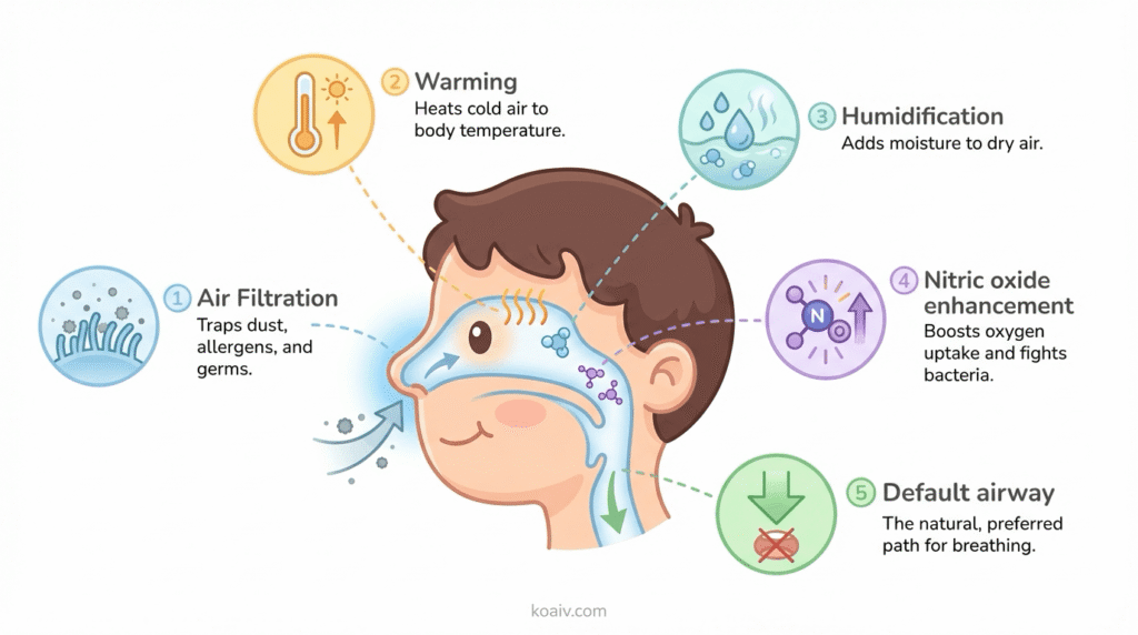
The nose is, in biological reality, the body’s most sophisticated air-processing system—a device that performs at least four critical functions on every single breath, all of them invisible, all of them essential.
Filtering: The First Line of Defense
Imagine walking through a construction site without a dust mask. Fine particles of debris, concrete powder, and airborne bacteria would flood directly into the throat and lungs. The nose functions as the body’s built-in dust mask. The nasal cavity is lined with tiny hairs called cilia (think of them as a microscopic forest of bristles) and coated with a layer of sticky mucus. Together, they form a filtration system that traps airborne particles—dust, pollen, bacteria, viruses, mold spores—before they can travel deeper into the respiratory system. The trapped material is then swept toward the throat by the rhythmic, wave-like beating of the cilia and swallowed, where stomach acid neutralizes it.
When a child breathes through the mouth, this entire filtration system is bypassed. Unfiltered air pours directly past the tonsils and into the throat and lungs—the biological equivalent of removing the air filter from a car engine and hoping for the best.
Warming and Humidifying: Climate Control in Milliseconds
The lungs are delicate organs. They function best when the air arriving at their surface is warm (close to body temperature, around 37°C / 98.6°F) and humid (near 100% relative humidity). Cold, dry air irritates the bronchial lining, triggers mucus overproduction, and can even provoke bronchospasm—the tightening of the airways that asthmatics know intimately.
The nasal cavity handles this with remarkable precision. Inside the nose are structures called turbinates—three sets of bony shelves on each side, covered in a rich blood supply and moist tissue. As air flows over and around these turbinates, it is warmed and humidified in a fraction of a second. Think of the turbinates as a radiator and a humidifier combined, operating automatically and instantaneously, calibrating themselves to the temperature and humidity of the incoming air.
When air enters through the mouth instead, it arrives at the lungs still relatively cold and dry—a suboptimal condition that the lower airways must then compensate for, at a cost to tissue health over time.
Nitric Oxide: The Secret Ingredient Most People Have Never Heard Of
This is perhaps the most fascinating—and least known—function of nasal breathing. The paranasal sinuses (hollow, air-filled cavities surrounding the nose) produce a gas called nitric oxide (NO). When a person breathes in through the nose, this gas is carried with the inhaled air down into the lungs.
What does nitric oxide do? It acts as a vasodilator—it relaxes and widens the blood vessels in the lungs. Wider blood vessels mean more blood flows past the air sacs (alveoli) where oxygen exchange occurs, and more oxygen is transferred into the bloodstream per breath. A study published in Acta Physiologica Scandinavicademonstrated that nasal breathing can increase oxygen absorption by 10 to 25%compared to mouth breathing of the same volume of air.
To put this in everyday terms: imagine two identical cars driving up the same hill—but one is running on premium fuel and the other on a watered-down blend. Both cars make it up the hill, but one does it far more efficiently. Nasal breathing is the premium fuel. The body can operate on mouth-breathed air, but it is working harder to extract less oxygen.
For a growing child—whose brain is consuming a disproportionate share of the body’s oxygen supply as it wires billions of new neural connections—this efficiency gap is not trivial.
The Big Picture
Here is a simple way to visualize the difference:
| Function | Nasal Breathing | Mouth Breathing |
|---|---|---|
| Air Filtration | ✅ Particles trapped by cilia and mucus | ❌ Unfiltered air reaches lungs directly |
| Air Warming | ✅ Warmed to body temperature via turbinates | ❌ Cool, dry air irritates airways |
| Humidification | ✅ Moistened to ~100% humidity | ❌ Dry air dries out throat and lung tissue |
| Nitric Oxide Boost | ✅ NO enhances oxygen uptake by 10–25% | ❌ No NO production; reduced oxygen efficiency |
| Default Airway | ✅ Evolutionary design; optimal pathway | ❌ Emergency backup; compensatory pathway |
The nose, in short, is not optional equipment. It is the body’s primary and preferred airway—refined over hundreds of thousands of years of human evolution. The mouth can breathe, certainly. But it was designed primarily for eating and speaking, not for breathing. When it is recruited for the latter function on a chronic basis, the body begins to pay a price—and in a growing child, that price is steeper than most people imagine.
This naturally raises a question: if nasal breathing is the body’s default, what causes a child to abandon it?
Why Children Become Mouth Breathers
Chronic mouth breathing is not a choice. No child wakes up one morning and decides to breathe through the wrong opening. It develops because something—structural, allergic, environmental, or habitual—has made nasal breathing difficult, uncomfortable, or impossible.
Understanding the root cause is not academic trivia; it is the essential first step in any treatment plan. The intervention for a child with massively enlarged adenoids is fundamentally different from the intervention for a child with chronic allergies or a child who has simply been breathing through the mouth so long that the habit has hardened into default.
The Anatomical Roadblocks
The most common structural cause of chronic mouth breathing in children involves two small but powerful pieces of tissue: the adenoids and the tonsils.
The adenoids are a pad of lymphatic (immune) tissue located high in the throat, directly behind the nasal passage. They are not visible by simply looking inside a child’s mouth—they sit in a hidden location where the back of the nose meets the top of the throat. Their job is to help fight infection by trapping pathogens that enter through the nose. In many children, however, the adenoids become chronically enlarged—either due to repeated infections, allergic inflammation, or simple genetic tendency. When they swell, they can physically block the nasal airway from behind, like a cork wedged into the back of a pipe. Air simply cannot pass through. The child’s only option is to open the mouth.
The tonsils—the two masses of tissue visible on either side of the back of the throat—can enlarge for similar reasons. When both the tonsils and adenoids are swollen, the combined obstruction can be significant, particularly in the narrow airway of a young child.
Dr. Christian Guilleminault, a pioneering sleep researcher at Stanford University who spent decades studying pediatric sleep-disordered breathing, identified adenotonsillar hypertrophy (enlarged adenoids and tonsils) as the single most common initiating cause of chronic mouth breathing and pediatric obstructive sleep apnea.
Other structural causes include a deviated nasal septum (the thin wall between the two nostrils is crooked, reducing airflow on one or both sides), nasal polyps (soft, painless growths inside the nasal passages), and tongue-tie (ankyloglossia)—a condition that deserves its own moment of explanation.
Tongue-tie occurs when the frenulum—the small band of tissue connecting the underside of the tongue to the floor of the mouth—is unusually short, thick, or tight. This restricts the tongue’s range of motion. Why does this matter for breathing? Because when the mouth is closed and a person is breathing nasally, the tongue naturally rests with its entire upper surface pressed gently against the roof of the mouth (the palate). This is the proper “resting posture” of the tongue.
A tongue-tied child often cannot achieve this position comfortably. The tongue drops to the floor of the mouth, the jaw opens to compensate, and mouth breathing follows. The significance of tongue posture will become even clearer in the next section.
Allergic and Environmental Triggers
Not all nasal obstruction involves oversized tissue. Chronic inflammation—swelling of the nasal lining itself—can be equally effective at blocking the airway.
Allergic rhinitis is the leading cause of this type of obstruction. When a child is allergic to dust mites, pet dander, mold, cockroach proteins, or pollen, the immune system treats these harmless substances as threats. It floods the nasal lining with inflammatory chemicals—histamine chief among them—causing the tissue to swell, produce excess mucus, and obstruct airflow. The child’s nose becomes perpetually stuffy, and the mouth opens as a compensatory reflex.
The scale of this issue is significant. The American Academy of Allergy, Asthma, and Immunology estimates that allergic rhinitis affects approximately 40% of children in developed nations. For many of these children, the congestion is not episodic but chronic—a constant low-grade inflammation that keeps the nasal passages perpetually swollen and underperforming.
Environmental pollutants act through a similar mechanism. Chronic exposure to cigarette smoke, vehicle exhaust, industrial pollutants, or even the volatile organic compounds (VOCs) emitted by new furniture, paint, and cleaning products can inflame the nasal mucosa and contribute to nasal obstruction.
The Habit That Outlives Its Cause
Perhaps the most surprising entry in this list is simple habit. The human brain is an adaptation machine. It is designed to find the most efficient behavioral pattern for a given situation and then automate it, freeing conscious attention for other tasks.
When a child spends an extended period breathing through the mouth—say, during a winter of back-to-back respiratory infections, or a season of severe allergies—the brain begins to encode mouth breathing as the default respiratory pattern. The neural pathways for nasal breathing grow weaker through disuse, while the mouth-breathing pathways strengthen.
The troubling result: even after the original obstruction resolves—the infections clear, the adenoids shrink, the allergy season ends—the mouth-breathing pattern can persist. The child’s nasal airway is now open, but the brain has stopped trying to use it. The habit has become self-sustaining, like a detour that was used so often it became the main road.
This is why identifying and addressing chronic mouth breathing early matters so much. The longer the pattern persists, the more deeply it embeds itself—not just in the nervous system, but, as the next section reveals, in the very bones of the face.
The Domino Effect: How the Wrong Breath Reshapes a Growing Child
The consequences of chronic mouth breathing extend far beyond the airway itself. They ripple outward in a cascading chain of effects—touching the architecture of the face, the depth of sleep, the sharpness of the mind, and the resilience of the immune system.
How Breathing Sculpts the Face
This is the consequence that surprises parents most—and for good reason. The idea that the simple act of breathing can reshape the bones of a child’s face sounds, on first hearing, implausible. It is not. It is one of the most well-documented phenomena in craniofacial biology.
The mechanism hinges on a principle that is easy to grasp once explained: during childhood, the bones of the face and jaw are not yet fully hardened. They are still growing, still responsive to the forces applied to them. Among the most important of those forces is the pressure of the tongue.
Picture a child breathing normally through the nose, mouth closed. The tongue, with nowhere else to go, rests with its broad upper surface pressed against the palate—the roof of the mouth. This is not a conscious action; it is the default resting posture of the tongue in a nasal breather. And that gentle, sustained pressure matters enormously. The palate is also the floor of the nasal cavity. When the tongue pushes outward against it, hour after hour, day after day, it encourages the upper jaw (maxilla) to grow wide and broad—creating a generous, U-shaped dental arch with plenty of room for all the permanent teeth to emerge in proper alignment.
Now picture the mouth-breathing child. The mouth is open, the jaw drops, and the tongue—deprived of its natural resting position against the palate—falls to the floor of the mouth. The outward pressure disappears. In its absence, the muscles of the cheeks—which are now the dominant force—push inward on the upper jaw. Over months and years, the palate narrows. It rises into a high, narrow, V-shaped arch. There is insufficient space for the permanent teeth. They crowd, overlap, and erupt crookedly.
But the changes do not stop at the teeth. The entire pattern of facial growth shifts. Without the tongue’s forward and outward pressure, the midface (the area around the cheekbones and nose) becomes flattened and underdeveloped. The lower jaw recedes. The face elongates vertically. Craniofacial researchers have a clinical name for this constellation of features: “adenoid facies” or “long-face syndrome.” The features include:
- A narrow, elongated face
- A recessed or “weak” chin
- An open-mouth resting posture
- Visible upper gums when smiling (a “gummy smile”)
- Flat cheekbones and underdeveloped midface
- Dark circles under the eyes
A 2015 study published in the European Journal of Orthodontics compared the craniofacial measurements of children identified as chronic mouth breathers to those of habitual nasal breathers. The differences were statistically significant and measurable—critically—as early as age 6. By the time most families notice the problem or seek treatment, the face has already been remodeling itself for years.
This is why pediatric dentists and orthodontists increasingly view the airway as the starting point of orthodontic assessment. Straightening teeth without addressing the underlying breathing pattern is, in many cases, treating the symptom while the cause continues its work in the background.
Sleep: The Hidden Casualty
If the facial changes represent the most visible consequence of chronic mouth breathing, the sleep disruption is arguably the most consequential—because sleep is not merely rest. For a child, sleep is when the most critical construction work of development occurs.
During deep sleep, the brain consolidates memories, processes the day’s learning, prunes unnecessary neural connections, and strengthens important ones. The pituitary gland releases growth hormone—the chemical signal that drives physical growth, bone development, and tissue repair. The immune system performs maintenance and surveillance. Disrupting this process is not like missing a meal; it is like pulling workers off a construction site while the building is still going up.
Chronic mouth breathing disrupts sleep through a direct mechanical pathway. When the mouth is open and the tongue is positioned low during sleep, the soft tissues of the throat—the soft palate, the uvula, and the base of the tongue—lose structural support and are more likely to vibrate (causing snoring) or collapse inward (causing obstruction). Each time the airway narrows or closes, the brain must rouse itself just enough to restore muscle tone and reopen the passage. These are called micro-arousals—brief surges of brain activity lasting only seconds, too short for the child to consciously remember, but long enough to shatter the continuity of deep sleep.
In mild cases, this produces restless, noisy sleep and a child who seems chronically “not quite rested.” In more severe cases, it constitutes pediatric obstructive sleep apnea (OSA)—a condition in which the airway fully closes repeatedly throughout the night, causing measurable drops in blood-oxygen levels between obstruction events.
The American Academy of Pediatrics estimates that 1 to 5% of children meet the clinical criteria for obstructive sleep apnea. But this figure captures only the tip of the iceberg. The broader category of sleep-disordered breathing—which includes habitual snoring, upper airway resistance syndrome, and subclinical obstruction—likely affects a far larger population of children, many of whom have never been evaluated.
The Misdiagnosis Trap: When an Airway Problem Looks Like a Behavior Problem
This is, in many ways, the most urgent message in the entire discussion—and the area where awareness could do the most immediate good.
A child who sleeps poorly does not always look sleepy. This is one of the least intuitive facts in pediatric medicine, and it catches countless parents and even some clinicians off guard. Adults who are sleep-deprived become sluggish, drowsy, and slow. Childrenwho are sleep-deprived often respond in the opposite direction: they become hyperactive.
The neuroscience behind this is still being fully mapped, but the prevailing theory is straightforward. When a child’s prefrontal cortex—the brain region responsible for impulse control, focus, and emotional regulation—is impaired by inadequate deep sleep, the child loses the internal “brakes” that normally modulate behavior. Without those brakes, the child becomes impulsive, distractible, emotionally volatile, and physically restless. In a word: hyperactive.
These symptoms—hyperactivity, inattention, impulsivity, emotional dysregulation—are the exact diagnostic criteria for Attention Deficit Hyperactivity Disorder (ADHD).
A landmark study published in the journal Pediatrics followed over 11,000 children and found that those with sleep-disordered breathing at ages 6 and 18 months were 40 to 100% more likely to develop behavioral problems resembling ADHD by age 7. Perhaps more striking, subsequent research has shown that when the underlying sleep-disordered breathing is treated—through adenotonsillectomy, myofunctional therapy, or other interventions—many children experience significant improvements in attention, behavior, and academic performance, sometimes to the degree that the ADHD-like symptoms resolve entirely.
This does not mean that ADHD is not a real condition. It is—and millions of children and adults live with it as a genuine neurodevelopmental difference. But the overlap between ADHD symptoms and sleep-deprivation symptoms is so substantial that a growing number of pediatric sleep specialists now advocate for airway and sleep evaluation as a standard component of any ADHD assessment. The question worth asking is not just “does this child have ADHD?” but also “is this child sleeping well enough for their brain to function at its baseline?”
The Body Under Siege: Immunity, Oral Health, and Posture
The consequences of chronic mouth breathing further extend into three domains that, while less dramatic, are no less important in aggregate.
Immune vulnerability. Every breath drawn through the mouth is a breath that bypasses the nasal filtration system. Bacteria, viruses, allergens, and particulates that would have been trapped by nasal cilia and mucus instead land directly on the tonsils, the pharynx, and the bronchial lining. Over time, this increased pathogen exposure translates into a higher incidence of sore throats, ear infections, upper respiratory infections, and even pneumonia. For many mouth-breathing children, the pattern creates a frustrating cycle: recurrent infections cause further swelling, which causes further obstruction, which reinforces mouth breathing.
Oral health decline. Saliva is not merely a digestive fluid. It is the mouth’s primary self-defense system—a complex solution containing antimicrobial enzymes, pH-buffering agents, and minerals (calcium and phosphate) that continuously repair and remineralize tooth enamel. When a child breathes chronically through the mouth, the constant airflow evaporates saliva from the teeth and gums. The mouth dries out. Without adequate saliva, cavity-causing bacteria multiply unchecked, the pH of the mouth drops into the acidic range where enamel dissolves, and the gums become inflamed. The result is a measurably higher rate of dental cavities, gingivitis, and chronic bad breath (halitosis) in mouth-breathing children—findings that have been consistently documented in the pediatric dental literature.
Postural compensation. A child struggling to move air through a compromised airway will instinctively do whatever it takes to open that airway further. One of the most common unconscious compensations is forward head posture—the chin juts forward, the neck extends, and the head tilts slightly upward. This position straightens and slightly widens the pharyngeal airway, making breathing marginally easier. But it does so at a cost: chronic strain on the muscles and joints of the cervical spine, tension headaches, rounded shoulders, and, over time, measurable changes in spinal alignment. Walk into any pediatric physical therapy clinic, and forward head posture associated with mouth breathing is a familiar presentation.
A Parent’s Guide to the Signs
Because chronic mouth breathing develops gradually—there is no sudden onset, no fever, no rash—it often escapes detection until its consequences are already well-established. This makes parental observation the most important early-detection tool available.
The following signs do not individually confirm chronic mouth breathing—many of them can be caused by other conditions. But when several cluster together, they form a pattern that warrants investigation.
What to Watch for at Night
- Audible breathing, snoring, or gasping. Any sound during sleep indicates turbulent airflow—air moving through a passage that is partially obstructed. Snoring in children is common but not normal. The American Academy of Pediatrics considers habitual snoring (more than three nights per week) a screening criterion for sleep-disordered breathing.
- Restless sleep and unusual positions. A child who tosses frequently, sleeps with the neck hyperextended (head tilted far back), or consistently sleeps propped up on pillows may be unconsciously positioning themselves to keep the airway open.
- Bedwetting (nocturnal enuresis). This connection surprises many parents. During obstructive sleep events, changes in chest pressure affect the heart, which releases a hormone called atrial natriuretic peptide (ANP). ANP signals the kidneys to produce more urine. At the same time, the micro-arousals from apnea events disrupt the child’s ability to sense a full bladder and wake. The result: bedwetting in a child who may have been dry for years. Studies have shown that treating sleep apnea resolves bedwetting in a significant proportion of affected children.
- Mouth open during deep sleep. This one seems obvious, but it is worth stating explicitly: if a child’s mouth is consistently open during deep sleep (not just during a cold), it is the most direct visual indicator of oral breathing.
What to Watch for During the Day
- Chronically dry, cracked lips. Constant airflow over the lips strips away moisture. Many mouth-breathing children develop a habit of licking their lips, which paradoxically worsens the dryness, creating a reddened, irritated ring around the mouth called “lip-licker’s dermatitis.”
- Dark circles under the eyes. Sometimes called “allergic shiners,” these are not caused by lack of sleep per se but by venous congestion—pooling of blood in the small veins beneath the eyes due to chronic nasal obstruction. They give the child a tired, hollow-eyed appearance even when the child has technically slept a full night.
- Open-mouth resting posture. Observe the child during quiet, focused activities—watching a screen, reading, doing homework. A chronically open mouth during these moments, when the body is at rest and nasal breathing should be effortless, is a significant indicator.
- Picky or slow eating. This is a link many parents miss. Chewing food requires the mouth to close. A child who is dependent on the mouth for breathing faces an uncomfortable conflict: close the mouth to chew, and breathing becomes labored. Many such children eat slowly, chew with their mouths open, prefer soft foods that require less chewing, or simply become “picky” eaters as a way of avoiding the discomfort.
- Difficulty concentrating, irritability, or hyperactivity. As discussed, these may be downstream symptoms of fragmented sleep rather than primary behavioral issues.
A practical tip: If something seems off, use a smartphone to record a short video of the child sleeping—ideally capturing both the sound and a clear view of the face. This 60-second recording can be more informative to a pediatrician or ENT than any verbal description. It captures what words alone often fail to convey: the snoring pattern, the open mouth, the restless repositioning, and sometimes even visible pauses in breathing.
The Action Plan: Building the Right Team
One of the defining characteristics of chronic mouth breathing is that it sits at the intersection of multiple medical and dental specialties. No single practitioner sees the entire picture. Effective management almost always requires a multidisciplinary team—a concept that can feel intimidating to families but that, in practice, often involves just two or three coordinated professionals.
The Pediatrician: The Starting Point
The family pediatrician is the natural first contact—the person who can listen to parental concerns, perform an initial assessment, and generate referrals. It is worth noting that awareness of mouth breathing’s consequences is growing rapidly but is not yet universal in primary care training. Parents who raise the concern clearly and specifically—ideally armed with a sleep video—are more likely to receive a thorough response.
The ENT Specialist: Assessing the Hardware
An otolaryngologist (ear, nose, and throat specialist) evaluates the physical structures of the airway. Using a combination of physical examination and, in some cases, a flexible nasopharyngoscopy (a thin, painless camera threaded through the nose to visualize the adenoids), the ENT can determine whether enlarged adenoids, tonsils, a deviated septum, or nasal polyps are contributing to the obstruction.
When enlarged adenoids and tonsils are confirmed as the primary cause, adenotonsillectomy (surgical removal of the adenoids and tonsils) is one of the most effective interventions available. A large-scale study called the CHAT trial (Childhood Adenotonsillectomy Trial), published in the New England Journal of Medicine, randomized children with obstructive sleep apnea to either surgery or watchful waiting. The surgical group showed significant improvements in sleep quality, behavior, and quality of life at the seven-month follow-up.
The Pediatric Dentist or Orthodontist: Guiding the Bones
A pediatric dentist or orthodontist evaluates the dental and skeletal consequences of mouth breathing—the narrow palate, the crowded teeth, the crossbite—and can intervene to redirect facial growth while the child is still growing.
One of the most powerful tools in this context is the palatal expander—a fixed or removable appliance that sits against the palate and applies gentle, continuous outward pressure to the midpalatal suture (the growth seam running along the center of the roof of the mouth). Over weeks to months, this suture gradually separates and new bone fills in, widening the upper jaw. The effect is twofold: the dental arch widens, creating room for the teeth, and the floor of the nasal cavity—which is, remember, the same structure as the palate—also widens, physically increasing the volume of the nasal airway.
The critical detail is timing. The midpalatal suture remains responsive to expansion until it begins to fuse, which typically occurs between ages 12 and 16. Intervention before this window closes is dramatically more effective and less invasive than intervention after.
The Orofacial Myofunctional Therapist: Retraining the Software
If the ENT addresses the hardware (removing structural obstructions) and the orthodontist guides the bones (expanding the palate), the orofacial myofunctional therapist (OMT) addresses the software—the neuromuscular patterns that govern how a child breathes, swallows, and positions the tongue at rest.
This is a field that many parents—and indeed many healthcare providers—have not yet encountered, but it is gaining rapid traction as the research base grows.
Myofunctional therapy is, at its core, physical therapy for the muscles of the face, tongue, lips, and throat. Through a structured program of daily exercises—typically lasting 10 to 15 minutes and spanning 6 to 12 months—an OMT teaches the child (and the child’s nervous system) to:
- Breathe through the nose as the default, both awake and asleep.
- Rest the tongue against the palate in the correct posture.
- Swallow correctly (using the tongue rather than the cheeks and lips to propel food).
- Seal the lips at rest without conscious effort.
The exercises are typically playful and age-appropriate—tongue push-ups against the palate, lip seal holds, controlled breathing drills—and are designed to build the strength and coordination necessary for these patterns to become automatic.
A 2015 meta-analysis published in the journal Sleep reviewed multiple studies of myofunctional therapy in children and adults with obstructive sleep apnea. The findings were striking: myofunctional therapy reduced the severity of sleep apnea, as measured by the apnea-hypopnea index, by approximately 62% in children. A later systematic review published in Sleep Medicine Reviews confirmed that myofunctional therapy is an effective adjunct treatment, particularly when combined with other interventions like surgery or orthodontics.
What Families Can Do at Home
Professional treatment is the cornerstone, but the home environment plays a powerful supporting role—particularly in managing allergen exposure, maintaining nasal hygiene, and reinforcing the transition to nasal breathing.
Managing the Indoor Environment
For children whose mouth breathing is driven or worsened by allergic rhinitis, reducing the allergen load in the home—and especially the bedroom, where the child spends 8 to 12 hours each night—can meaningfully reduce nasal inflammation and improve airway patency.
Evidence-supported strategies include:
- HEPA air purifiers in the child’s bedroom (HEPA filters capture 99.97% of particles 0.3 microns and larger, including dust mite allergen, pollen, and mold spores).
- Allergen-proof encasings on the mattress, pillows, and duvet. Dust mites—the most common indoor allergen—live primarily in bedding.
- Washing bedding weekly in hot water (at least 60°C / 140°F) to kill dust mites.
- Removing or reducing carpet in the bedroom (hard flooring harbors far fewer allergens).
- Keeping pets out of the bedroom (even if the child is not formally allergic, pet dander is a potent nasal irritant).
Nasal Saline Irrigation
Nasal saline rinses—using a gentle saline spray, a squeeze bottle, or a neti pot adapted for children—have a robust evidence base for reducing nasal congestion and improving airway clearance. A Cochrane review found that saline irrigation is a safe, inexpensive, and effective intervention for both allergic and non-allergic rhinitis in children.
The mechanism is straightforward: the saline solution physically flushes mucus, allergens, and inflammatory mediators from the nasal passages, reducing swelling and restoring airflow. Most children over the age of 3 or 4 can learn to tolerate and even enjoy nasal rinsing when it is introduced patiently and playfully.
Breathing Awareness Through Play
Nasal breathing awareness can be reinforced during the day through simple, enjoyable activities:
- Bubble blowing. Blowing bubbles requires exhaling through the mouth but inhaling through the nose (the mouth is occupied by the wand). It is a natural nasal-breathing trainer disguised as play.
- Pinwheel spinning. Similar principle—controlled exhalation through the mouth, nasal inhalation.
- Humming games. Humming can only be done with a closed mouth and nasal airflow. It also happens to stimulate nitric oxide production in the sinuses.
- Sticker or feather exercises. Holding a thin sticker or small feather between closed lips during a quiet activity (watching a show, coloring) builds lip-seal endurance in a low-pressure way.
A Critical Safety Note
The practice of mouth taping—applying adhesive tape over the lips during sleep to force nasal breathing—has gained significant visibility through social media and wellness communities. For children, this practice carries serious safety risks and should never be attempted without explicit guidance from a medical professional who has confirmed that the child’s nasal airway is fully patent. A child with any degree of nasal obstruction who is taped into mouth closure could experience dangerous oxygen desaturation. The risks dramatically outweigh any potential benefit when attempted outside of medical supervision.
Conclusion
There is a concept in architecture that form follows function—that the shape of a structure should emerge from its purpose. The same principle operates, with quiet precision, in the human body. The way a child breathes—day after day, year after year, through thousands of sleep cycles and millions of individual breaths—shapes the architecture of the face, the quality of the sleep, the efficiency of the oxygen supply, and, downstream, the capacity for attention, learning, and emotional regulation.
Chronic mouth breathing is not a childhood phase that resolves on its own. It is a physiological pattern with measurable, evidence-backed consequences—consequences that are most reversible when addressed during the window of craniofacial growth and neural plasticity that childhood provides. Once the midpalatal suture fuses, once the facial bones harden into their adult form, once the neural pathways for mouth breathing have been reinforced for a decade or more, intervention becomes more complex, more invasive, and less predictable.
The encouraging counterpart to that urgency is this: the interventions work. Adenotonsillectomy can restore nasal airway patency overnight. Palatal expansion can reshape the jaw and widen the nasal floor in a matter of months. Myofunctional therapy can retrain the neuromuscular patterns of a lifetime in under a year. Allergy management and environmental controls can reduce the inflammatory burden on the nasal passages within weeks. And the downstream effects of these interventions—better sleep, sharper focus, healthier teeth, a face that develops to its genetic potential—compound over a lifetime.
It all starts with a single act of noticing. With pausing in that doorway, watching a sleeping child, and asking a question that is simple, powerful, and too rarely asked:
Should a perfectly healthy child be breathing through the mouth?
In almost every case, the answer is no. And in almost every case, something can be done about it.
References & Further Reading